Cell: The Fundamental Unit of Life

Cell: The FundamentalnUnit of Life - Class 9 Science

  • Brief History of Cell
  • Characteristics of Cell
  • Shape and Size
  • Components of a Cell
  • Solved Questions on Cell- The Fundamental Unit of Life
  • Brief History of Cell

    The discovery of cells marked a significant milestone in the history of biology.

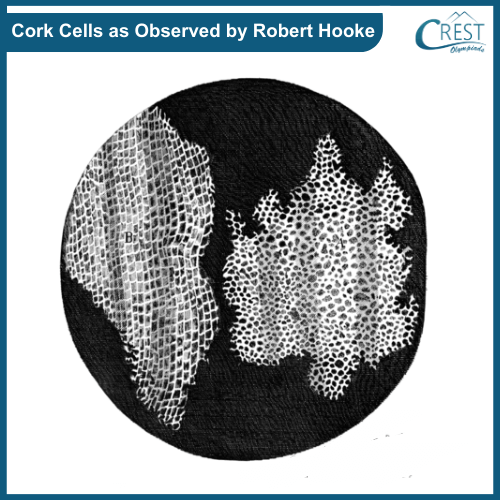
    Robert Hooke (1665): Robert Hooke, an English scientist, used a primitive microscope to examine slices of cork and other materials. He coined the term "cell" to describe the small compartments he observed in cork, which reminded him of the cells in a monastery.

    Hooke's observations of cork cells were groundbreaking, as they revealed the existence of small structural units in living organisms that had not been recognized before. However, it's important to note that Hooke's microscope was not powerful enough to reveal the complexity of living cells.

    Cork Cell - Cell Observed by Robert Hooke

    Anton van Leeuwenhoek (Late 17th Century): Around the same time, the Dutch scientist Anton van Leeuwenhoek achieved higher magnification in his microscopes and made detailed observations of various microscopic organisms, including bacteria and protozoa. He is often considered the "Father of Microbiology" for his pioneering work in microscopy.

    Matthias Schleiden and Theodor Schwann (19th Century): The German botanist Matthias Schleiden and the German physiologist Theodor Schwann formulated the cell theory in the 19th century. Schleiden concluded that all plants are composed of cells, and Schwann extended this idea to animals. The cell theory states that:

    1. All living organisms are composed of one or more cells.
    2. The cell is the basic unit of structure, function, and organization in all organisms.
    3. All cells arise from pre-existing cells.

    Rudolf Virchow (1855): The German physician Rudolf Virchow contributed to the cell theory by emphasizing the idea of cell division and stating "omnis cellula e cellula," which means "every cell originates from another existing cell." This concept further solidified the understanding that cells are the fundamental units of life.

    Characteristics of Cell

    1. Cells as Building Blocks: Cells are the fundamental units of all living organisms, serving as the building blocks of life.

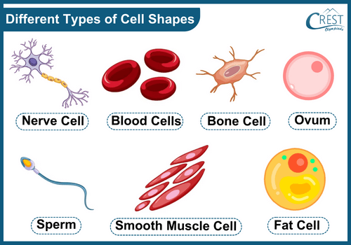
    2. Variation in Cells: Cells vary in terms of shape, size, and functions. Different types of cells perform specialized roles within an organism.

    3. Unicellular Organisms: Some organisms consist of a single cell that carries out all life processes. Examples include Chlamydomonas, paramecium, and bacteria.

    4. Specific Functions: Each living cell has specific functions that contribute to the overall functioning of the organism.

    5. Organelles: Cells share common organelles that carry out essential functions, although there may be variations between animal and plant cells.

     Learn about Reproduction in Organisms

    Shape and Size

    1. Function Determines Shape and Size: The shape and size of cells are influenced by the specific functions they perform within an organism.

    2. Unicellular Organisms: In unicellular organisms, a single cell carries out a variety of functions like digestion and locomotion. The cell's shape and size are adapted to facilitate these functions.

    3. Multicellular Organisms: In multicellular organisms, different types of cells have distinct shapes, sizes, and internal compositions to fulfil specialised functions. For example, muscle cells, nerve cells, and sperm cells each have unique features tailored to their roles.

    Different Types of Cell Shapes - Definition, Characterstics etc

    Components of a Cell

    1. Plasma Membrane

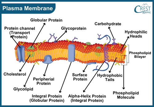
    The plasma membrane is the protective outer covering of a cell that defines its boundary and separates the cell from its external environment.

    Structure of Plasma Membrane

    Composition: The plasma membrane is primarily composed of lipids (such as phospholipids) and proteins. These components are arranged in a bilayer structure, with the hydrophilic (water-attracting) heads of the lipids facing outward and the hydrophobic (water-repelling) tails facing inward.

    Protein Distribution: Proteins are scattered throughout the lipid bilayer, and they have various roles including the transportation of molecules, cell signalling, and structural support.

    Synthesis: The smooth endoplasmic reticulum is involved in the synthesis of lipids, including those used in building the cell membrane. This process is known as membrane biogenesis.

    Structure of Plasma Membrane - Components of a Cell

    Functions of Plasma Membrane

    a) Cell Boundary: It defines the cell's outer boundary, protecting and enclosing its contents.

    b) Selective Permeability: It controls the passage of substances in and out of the cell, maintaining a controlled internal environment.

    c) Transport: It aids in the movement of ions, nutrients, and waste between the cell and its surroundings.

    d) Endocytosis: It allows the cell to engulf external material, both solid and liquid, through vesicle formation. In phagocytosis, cells like Amoeba engulf solid particles, and in pinocytosis, they ingest liquid droplets.

    e) Cell Signaling: It receives signals from the environment and transmits them to the cell's interior.

    f) Maintaining Shape: Together with the cytoskeleton, it maintains the cell's shape and structure.

    g) Homeostasis: It helps regulate internal conditions to keep the cell's environment stable.

    Transport of Materials through Plasma Membrane

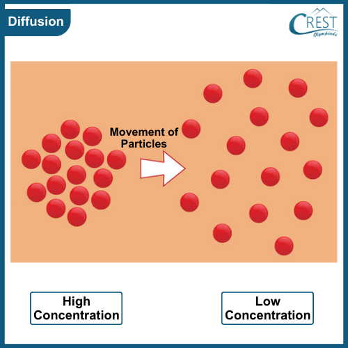
    Diffusion: Diffusion is the spontaneous movement of particles from an area of higher concentration to an area of lower concentration. It occurs due to random molecular motion and does not require energy. It plays a vital role in the exchange of gases, nutrients, and waste products within cells and between cells and their environment.

    Process of Diffusion - Transport of Materials through Plasma Membrane

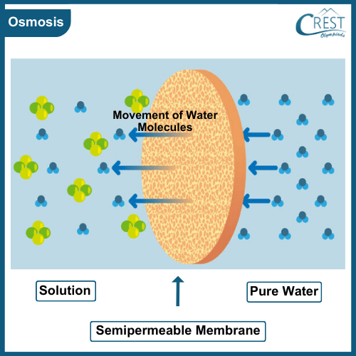
    Osmosis: Osmosis is a special type of diffusion involving the movement of water molecules across a semi-permeable membrane. Water moves from an area of lower solute concentration to an area of higher solute concentration. This process helps in maintaining the water balance within cells and is crucial for their survival.

    Process of Osmosis - Transport of Materials through Plasma Membrane

    Active Transport: Active transport is a process that moves molecules against their concentration gradient, i.e., from an area of lower concentration to an area of higher concentration. It requires energy (usually in the form of ATP) and involves specific protein pumps embedded in the plasma membrane. Active transport is essential for the uptake of essential nutrients and the removal of waste products, even when the concentration gradient goes against natural diffusion.

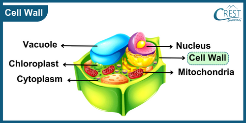
    2. Cell Wall

    The cell wall is a protective and structural layer that surrounds the plasma membrane of plant cells, fungi, bacteria, and some other organisms. It provides shape, support, and protection to the cell, much like the outer walls of a building.

    Composition

    The main component of the cell wall in plants is cellulose, a complex carbohydrate made up of long chains of glucose molecules. Cellulose fibres are arranged in a matrix, giving the cell wall its strength and rigidity. In addition to cellulose, the cell wall may also contain other carbohydrates, proteins, and substances like lignin, which further contribute to its strength.

    Structure of Cell Wall

    The cell wall is usually composed of multiple layers, each with different properties. The primary cell wall is the outermost layer and is relatively flexible. As the cell grows and matures, it may develop a secondary cell wall beneath the primary cell wall. The secondary cell wall is thicker, denser, and provides additional strength and protection.

    Diagram of Structure of Cell Wall - Science Grade 9

    Functions of Cell Wall

    a) Structural Support: The cell wall gives plants their characteristic shape and rigidity. It helps the plant cells maintain their shape even in changing environmental conditions.

    b) Protection: The cell wall acts as a barrier, protecting the cell from physical damage, pathogens, and other external threats. It acts as the first line of defence against mechanical stress, pathogens, and herbivores.

    c) Regulation of Water Uptake: The cell wall controls the movement of water into and out of the cell. It helps prevent excessive water uptake in hypotonic environments, preventing the cell from bursting due to osmotic pressure.

    d) Support in Plant Growth: As plant cells expand and grow, the cell wall provides resistance against the turgor pressure generated by the influx of water. This pressure is essential for maintaining the stiffness of the plant.

    e) Communication: The cell wall contains various signalling molecules that play a role in cell-to-cell communication, growth, and development.

    f) Transport: Although not as permeable as the plasma membrane, the cell wall allows for the exchange of certain substances between neighbouring cells.

    Important Terms

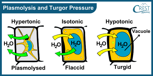
    Plasmolysis: When a plant cell is placed in a solution with a higher concentration of solutes than the cell's contents, water moves out of the cell through osmosis. As a result, the cell's contents shrink and pull away from the cell wall. This is called plasmolysis. The cell wall helps maintain the structural integrity of the cell even during plasmolysis, preventing the cell from collapsing.

    Turgor Pressure: Turgor pressure is the pressure exerted by the cell contents against the cell wall when the cell takes in water. The cell wall's rigidity resists excessive expansion of the cell, creating pressure that gives the plant cell its firmness and shape. Turgor pressure is crucial for maintaining the overall structure of the plant and supporting various physiological processes like nutrient transport and growth.

    Plasmolysis and turgor Pressure - Science Grade 9

    3. Nucleus

    Structure and Location

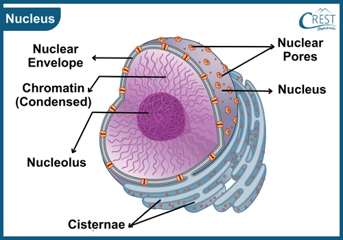
    The nucleus is a crucial cellular component found at the centre of eukaryotic cells. It typically takes on a spherical shape and is surrounded by a double-layered protective membrane called the nuclear membrane. This membrane serves as a barrier between the nucleus and the rest of the cell.

    Components Within the Nucleus

    a) Nuclear Membrane: This double-layered membrane encloses the nucleus, effectively separating its contents from the cytoplasm. It is studded with tiny pores that act as gateways, enabling the controlled movement of molecules between the nucleus and the cytoplasm.

    b) Nucleoplasm: Inside the nucleus, the nucleoplasm is a semi-fluid substance that houses various cellular components. It provides the medium in which critical nuclear processes take place.

    c) Nucleolus: The nucleolus is a distinct structure within the nucleus, often visible as a darker region.

    Labelled Diagram of Nucleus - Science Grade 9

    Distinction Between Prokaryotes and Eukaryotes

    Cells can be classified into two categories based on their nucleus structure: prokaryotes and eukaryotes.

    1. Prokaryotic cells, such as bacteria, lack a well-defined nucleus and nuclear membrane. Their genetic material is located in a region called the nucleoid.
    2. Eukaryotic cells, found in plants, animals, fungi, and protists, possess a membrane-bound nucleus that contains the genetic material.

    Characteristic

    Prokaryotes

    Eukaryotes

    Cell size

    Small (Generally 1-10 micrometres)

    Large (5-100 micrometres)

    Nucleus

    Not well-defined; lacks nuclear membrane and nucleolus

    Well-defined nucleus with nuclear membrane, nucleoplasm

    Number of Chromosomes

    Typically one circular chromosome

    Multiple linear chromosomes

    Nucleolus

    Absent

    Present

    Cell Organelles

    Few and simple organelles

    Membrane-bound organelles such as mitochondria, plastids, etc.

    Cell Division

    Binary fission (asexual)

    Mitosis (asexual) and Meiosis (sexual)

    Examples

    Bacteria and Archaea

    Plants, Animals, Fungi, and Protists

    Functions of Nucleus

    a) Genetic Information and Chromosomes: One of the nucleus's most vital functions is storing and safeguarding the cell's genetic information. This information is stored within structures known as chromosomes. Chromosomes consist of long DNA molecules wrapped around proteins. Each species has a specific number of chromosomes, and humans have 46.

    b) DNA and Gene Expression: DNA, or deoxyribonucleic acid, is the carrier of genetic information. Genes are specific segments of DNA that contain the instructions for creating proteins and guiding various cellular functions. These instructions are encoded in the sequence of nucleotides along the DNA molecule.

    c) Control and Regulation: Genes serve as the control switches for the cell's activities. They dictate how proteins are synthesised, determining the cell's behaviour, structure, and responses to environmental cues. This process is critical for the growth, development, and proper functioning of the organism.

    d) Cellular Reproduction: The nucleus plays a central role in cellular reproduction, the process by which a single cell divides and gives rise to new cells.

    e) Cell Development and Differentiation: The nucleus plays a crucial role in determining how a cell will develop and what form it will take on.

    f) Inheritance: The nucleus contains the genetic information that is passed from one generation to the next. During sexual reproduction, the genetic material from both parents is combined in the nucleus of the offspring, leading to genetic diversity within a population.

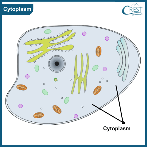
    4. Cytoplasm

    Cytoplasm is a vital component of a cell that plays a fundamental role in supporting various cellular activities. It is a gel-like, semi-fluid substance that fills the space between the cell's plasma membrane (outer boundary) and the nuclear membrane (if present). The cytoplasm contains various organelles, molecules, and structures that are essential for the cell's survival and functioning.

    Characteristics of Cytoplasm

    a) Composition: Cytoplasm is composed of water, ions, organic molecules, and various cellular structures such as organelles.
    b) Consistency: It has a gel-like consistency that provides support to the organelles and other cellular components.
    c) Motion: Some organelles within the cytoplasm, such as mitochondria and vesicles, can move within the cytoplasm through a process called cytoplasmic streaming or cyclosis.

    Diagram of Cytoplasm - Definition and Characterstics

    Functions of Cytoplasm

    a) Site of Cellular Activities: Many important cellular processes occur within the cytoplasm. Metabolic reactions, such as glycolysis (sugar breakdown) and protein synthesis, take place in this region.

    b) Support for Organelles: The cytoplasm provides a medium for organelles to be suspended in and maintain their positions. This organization is crucial for the proper functioning of organelles and their interactions.

    c) Transport: Molecules, nutrients, and waste products move within the cytoplasm, facilitating the transport of materials between different cellular compartments and organelles.

    d) Storage of Nutrients and Waste: The cytoplasm can store certain nutrients, ions, and waste products that are either awaiting use or awaiting excretion from the cell.

    5. Cell Organelles

    1. Cell organelles are specialised, membrane-bound structures found within eukaryotic cells. Each organelle has a specific structure and function, contributing to the overall activities of the cell.
    2. These organelles are crucial for maintaining the cell's internal organization, performing metabolic processes, and ensuring its survival. Inside a cell, you can find various organelles such as the Golgi apparatus, endoplasmic reticulum, lysosomes, mitochondria, plastids, and vacuoles.

    I. Endoplasmic Reticulum (ER)

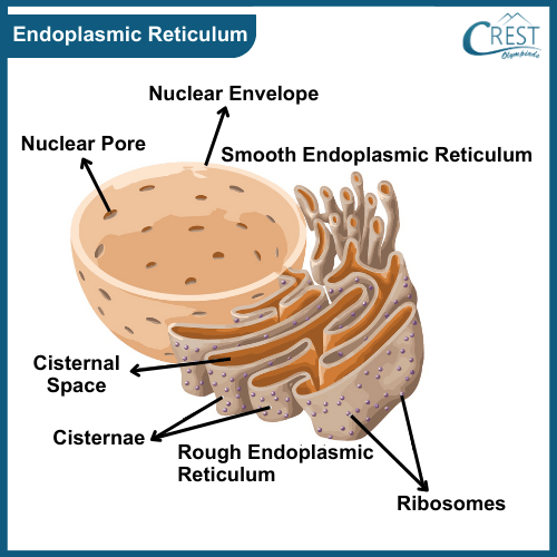
    The endoplasmic reticulum (ER) is an extensive network composed of membrane-bound tubules and sheets, serving as a transportation and distribution system for materials within the cell.

    There are two types of ER: rough endoplasmic reticulum (RER) and smooth endoplasmic reticulum (SER).

    Labelled Diagram of Endoplasmic Reticulum - Definition, Types and Function

    1. Rough Endoplasmic Reticulum (RER)

    1. The rough endoplasmic reticulum (RER) is a complex network of interconnected membrane-bound structures within a cell. Its distinctive rough appearance is due to the presence of ribosomes attached to its surface. Ribosomes are the cellular machinery responsible for protein synthesis.
    2. The RER serves as a crucial site for the synthesis, folding, and processing of proteins that are either secreted from the cell or used within the cell membrane.
    3. The RER plays a role in quality control, ensuring that proteins are properly folded and preventing misfolded or defective proteins from being transported further. It also aids in the synthesis of membrane proteins, which are important for the structure and function of the cell membrane.

    2. Smooth Endoplasmic Reticulum (SER)

    1. The smooth endoplasmic reticulum (SER) is a network of membranous structures within a cell that lacks ribosomes on its surface, giving it a smooth appearance when viewed under a microscope.
    2. The SER serves various functions in the cell, including the synthesis and distribution of lipids (fats) and steroids. It plays a crucial role in lipid metabolism, where it synthesizes lipids such as phospholipids and cholesterol, which are essential components of cell membranes.
    3. Additionally, the SER is involved in the detoxification of drugs and poisons in the liver cells by modifying them to make them more water-soluble and easier to eliminate from the body.
    4. The smooth endoplasmic reticulum also plays a role in storing calcium ions, which are important for various cellular processes, including muscle contraction and signal transduction.

    II. Golgi Apparatus

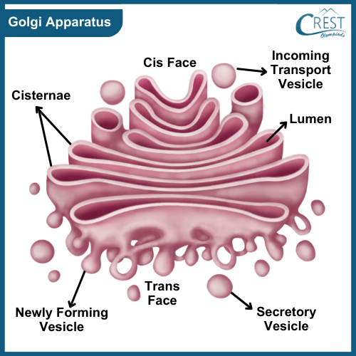
    The Golgi apparatus, often referred to as the Golgi complex or Golgi body, is a cellular organelle found in eukaryotic cells. It was named after its discoverer, Italian scientist Camillo Golgi.

    Structure of Golgi Apparatus

    The Golgi apparatus is composed of a series of flattened, membrane-bound sacs called cisternae. These cisternae are arranged in stacks that can vary in number from a few to many, depending on the cell type and its functions. Each stack has distinct regions, including the cis face (forming face), medial region, and trans face (maturing face).

    Labelled Diagram of Golgi Apparatus - Definition, Structure and Function

    Function of Golgi Apparatus

    The Golgi apparatus performs several essential functions in the cell:

    a) Modification and Processing: The Golgi apparatus is involved in modifying and processing various molecules, such as proteins and lipids, that are synthesized in the endoplasmic reticulum (ER).

    b) Protein Sorting: The Golgi acts as a molecular "post office," ensuring that different molecules are sent to the appropriate cellular compartments or secreted outside the cell.

    c) Packaging in Vesicles: The Golgi apparatus packages molecules into vesicles. These vesicles can carry modified proteins, lipids, or other molecules to various parts of the cell, such as the plasma membrane, lysosomes, or secretory vesicles for export.

    d) Formation of Lysosomes: The Golgi apparatus plays a crucial role in forming lysosomes. Enzymes that are required for digestion within lysosomes are synthesized in the ER, modified in the Golgi, and packaged into vesicles that become lysosomes.

     Explore more about Hereditary and Evolution

    II. Lysosomes

    Lysosomes are membrane-bound organelles found within the cells of eukaryotic organisms. These organelles play a vital role in cellular waste management, digestion, and various other cellular processes. Lysosomes are often referred to as the "waste disposal system" or the "cleanup crew" of the cell due to their function in breaking down and recycling cellular materials.

    Structure of Lysosomes

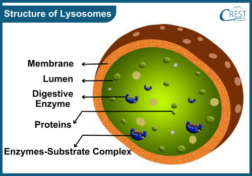
    Lysosomes are small, spherical organelles enclosed by a single lipid bilayer membrane. This membrane contains various proteins and enzymes that are responsible for the organelle's functions. The enzymes within lysosomes are typically acidic hydrolases, which are specialised for breaking down various types of molecules.

    Labelled Diagram of Structure of Lysosomes - Definition and Function

    Function of Lysosomes

    The primary functions of lysosomes include:

    a) Intracellular Digestion: Lysosomes contain a variety of enzymes capable of breaking down complex molecules, such as proteins, lipids, nucleic acids, and carbohydrates. These enzymes function optimally in the acidic environment within the lysosome. When cells engulf particles through processes like phagocytosis or endocytosis, lysosomes fuse with the vesicles containing these particles, releasing their enzymes to break down and digest the contents.

    b) Autophagy: Lysosomes are involved in autophagy, a process by which cells degrade and recycle their own damaged or unnecessary components. This is essential for maintaining cellular health and preventing the accumulation of malfunctioning structures.

    c) Phagocytosis: In immune cells, lysosomes are involved in phagocytosis, the process of engulfing and digesting foreign particles, such as bacteria or cellular debris.

    IV. Mitochondria

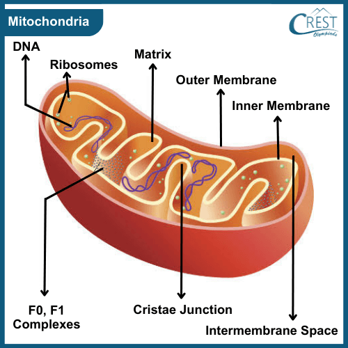
    Mitochondria are specialized organelles found within eukaryotic cells, which are the building blocks of complex organisms. These organelles are often referred to as the "powerhouses of the cell" due to their central role in producing energy that fuels various cellular activities.

    Structure of Mitochondria

    Outer Membrane: This is the outer boundary of the mitochondrion, separating it from the surrounding cellular environment. It contains transport proteins that allow the passage of certain molecules.

    Inner Membrane: The inner membrane is highly folded into structures called cristae. These folds increase the surface area available for chemical reactions. The inner membrane is where ATP (energy) synthesis takes place.

    Matrix: The interior of the mitochondrion, known as the matrix, contains enzymes, ribosomes, DNA, and other molecules necessary for energy production and other mitochondrial functions.

    Labelled Diagram of Mitochondria - Definition, Function etc

    Functions of Mitochondria

    a) Energy Production: The primary function of mitochondria is to generate energy in the form of adenosine triphosphate (ATP). ATP serves as a universal energy carrier that powers numerous biochemical processes within the cell, including muscle contraction, cellular division, and the synthesis of molecules needed for growth and maintenance.

    b) Cell maintenance: Mitochondria play a crucial role in cellular health, and their dysfunction is associated with various diseases and ageing processes. Disorders of mitochondria can lead to energy deficiencies and contribute to conditions such as mitochondrial diseases, neurodegenerative disorders, and metabolic disorders.

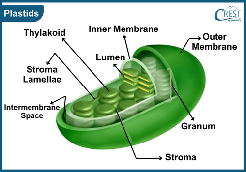
    V. Plastids

    Plastids are specialised organelles found in plant cells that play diverse roles in the life of a plant. These organelles are surrounded by a double-layered membrane and contain their own DNA and ribosomes. Plastids are involved in various essential functions within plant cells.

    Labelled Diagram of Plastids - Definition, Types and Functions etc

    Types of Plastids

    Plastids can be classified into different types based on their functions and contents:

    a) Chloroplasts: These are the most well-known type of plastids and are responsible for photosynthesis. Chloroplasts contain pigments like chlorophyll that capture light energy from the sun and convert it into chemical energy. This energy is used to synthesize glucose and other organic molecules from carbon dioxide and water.

    b) Chromoplasts: Chromoplasts are plastids that contain various pigments other than chlorophyll. These pigments give vibrant colours to flowers, fruits, and other plant parts, attracting pollinators and aiding in seed dispersal.

    c) Leucoplasts: Leucoplasts are colourless plastids that are involved in the storage of starch, oils, and proteins. They are important for storing energy reserves and nutrients within the cell.

    Functions of Plastids

    The main functions of plastids include:

    a) Photosynthesis: Chloroplasts are the primary sites of photosynthesis. They capture light energy and use it to produce glucose and oxygen from carbon dioxide and water. This process provides energy and organic molecules to the plant and releases oxygen into the atmosphere.

    b) Pigment Production: Plastids, especially chromoplasts, produce pigments that give colour to various plant structures. These pigments serve various purposes, from attracting pollinators to protecting plants from harmful ultraviolet (UV) radiation.

    c) Storage: Leucoplasts store starch, oils, and proteins. These stored substances can be used as reserves during times of energy or nutrient shortage

    VI. Vacuoles

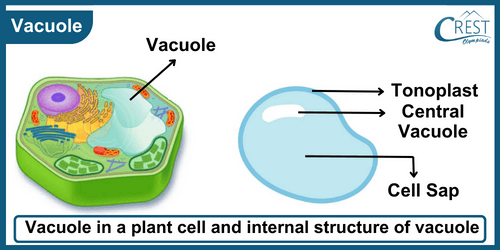
    Vacuoles are essential organelles found in both plant and animal cells, but they differ in size and function between these two types of organisms.

    a) In Animal Cells: In animal cells, vacuoles are generally smaller and more numerous. They are involved in storing various substances and aiding in cellular processes.

    b) In Plant Cells: In contrast, plant cells usually have fewer but much larger vacuoles that can occupy a significant portion of the cell's volume. These central vacuoles are mainly responsible for maintaining the plant's turgidity, storage, and waste disposal.

    Labelled Diagram of Vacuoles - Definition of Essential Organelles, Types etc

    Functions of Vacuoles

    The functions of vacuoles include:

    a) Turgor and Rigidity: In plant cells, vacuoles play a crucial role in maintaining turgor pressure, which gives the cell its rigidity and shape. When the central vacuole is filled with water (cell sap), it creates internal pressure against the cell wall, allowing the cell to maintain its structure.

    b) Storage: Vacuoles store a variety of substances, including water, ions, sugars, pigments, and even toxins. These stored compounds can be used for various cellular processes and can also act as reservoirs for nutrients and other essential molecules.

    c) Waste Disposal: Vacuoles function as a waste disposal system, storing harmful metabolic byproducts and toxins, and isolating them from the rest of the cell to prevent damage.

    d) Digestion: In some organisms like Amoeba, vacuoles are involved in the digestion of food. The process involves enclosing food particles within vacuoles and breaking them down using enzymes.

    e) Osmoregulation: In unicellular organisms, vacuoles help regulate the osmotic balance of the cell by expelling excess water or accumulating water to maintain a suitable internal environment.

    f) pH and Ion Balance: Vacuoles help regulate the pH and ion concentrations within the cell, contributing to overall cellular homeostasis.

    Share Your Feedback

    CREST Olympiads has launched this initiative to provide free reading and practice material. In order to make this content more useful, we solicit your feedback.

    Do share improvements at info@crestolympiads.com. Please mention the URL of the page and topic name with improvements needed. You may include screenshots, URLs of other sites, etc. which can help our Subject Experts to understand your suggestions easily.

    Other Science Related Topics Class 9

    70%